| back to AP home |
In 2006, the collaboration of 19 leading authorities led to a new estimate of amphibian phyologeny. It
was published in The Bulletin of the American Museum of Natural History(Volume 297, Issue 1, March,
2006). The title of the publication is The Amphibian Tree of Life. In this section of the website,
I planned to place scanning electron microscope images of APs from a fairly wide variety of anurans (frogs, toads
and their allies). They were part of a large study I carried out, beginning in the mid-1970s, to provide
inferences about AP evolution. The species were selected strategically on the basis of the generally
accepted estimates of phylogeny at that time. They were published largely in a journal (Scanning
Electron Microscopy) that is not widely available, so I thought it appropriate to place representative
examples here.
In the process of doing so, I learned of the 2006 work (through Wikipedia), and, on studying the new
tree of life realized that there were new tales to tell about the evolution of the anuran amphibian
papilla. The new estimate of phylogeny is based not only on morphological characters, but also on analyses
of mitochondrial and nuclear DNA. The base of the new tree, representing the anura with the most ancient
pedigrees, is similar to that of the old estimates of phylogeny, and is reasonably well covered by
my original selection of species. For the most ancient branch, the Leiopelmatidae, I have APs from two
of the four living species, including both genera (Ascaphus and Leiopelma). For the next
branch, the Xenoanura, I have APs from four species (two genera) from the Pipidae, one of two families
in the branch. For the third branch, the Costata, I have APs for two species from one family
(Bombinatoridae) and APs for one species from the other family (Alytidae). For the fourth branch,
Anomocoela, I have APs for species from two of the four families (from Pelobatidae and Scaphiopodidae).
At this point the new tree proceeds into the neobatrachia, the anurans of more modern origins. I
have no APs from species in the first neobatrachian branch, the Heleophrynidae- - the ghost frogs of
Southern Africa. If I were continuing the study today, that would be the first group of frogs I would go
for. The remaining neobatrachians have been grouped as the Phthanobatrachia, comprising two major limbs--
each of which needs to be climbed separately in our comparative morphology. For each limb, the Hyloides
and the Ranoides, I have APs from species at the first branch (Sooglossidae for Hyloides, Allodapanura for
Ranoides). For the Hyloides, I also have representative APs from an early middle branch, Brachycephalidae,
a later middle branch, Hylidae, and terminal, or near terminal branches, Dendrobatidae and Bufonidae.
For the Ranoides, I have no representatives from the middle branches, but I do have APs for species from
terminal or near-terminal branches, Rhacophoridae and Ranidae. If I were continuing the study today, I
would go for species in the mid-branches of the Ranoides limb (Ptychadenidae, Ceratobatrachidae,
Micrixalidae, Phrynobatrachia); but most excitedly, I would go for frogs in the Terrarana of Hedges et al.,
see APs on an Eleutherodactylus Tree
Ascaphus truei
|
Xenopus tropicalis
|
Alytes obstetricans
|
Pelobates fuscus
|

|
|
| 
|
| 
|
Sooglossus thomasetti
|
Gastrophryne olivacea
|
The Amphibian Papilla from Ancient to Modern Frogs and Toads
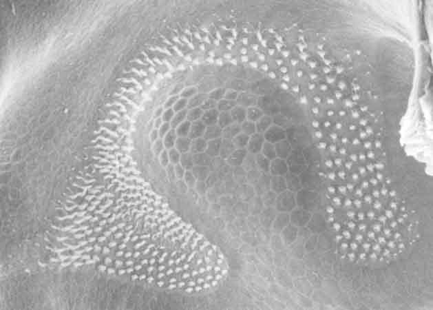
The AP in the upper left belongs to a member of the most ancient family (Ascaphidae or Leiopelmatidae)
of living anurans-- the tailed frog (Ascaphus truei). It consists of a single patch of sensory
epithelium. There are three other living members of this family. In the one that we examined, Leiopelma
hochstetteri, the AP also consisted of one patch. Next, in the upper row, is the AP from a species
(Xenopus tropicalis) in the branch nearest the Leiopelmatidae. This Xenopus AP consists of two patches,
which was true of all of the other anuran APs that we examined. In X. laevis, Cheuk Li observed the APs of
tadploes at various stages and found that the two patches were widely separated (by a field of squamous epithelial
cells) in early stages.
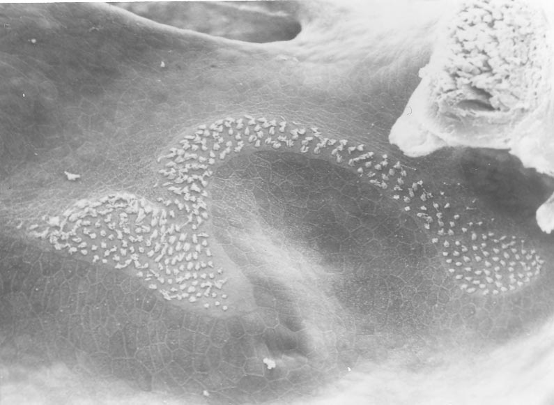
Next is the AP from the common midwife toad (Alytes obstetricans) a species from the third branch of the tree.
Immediately to the right of the Alytes AP is one from the common spadefoot (Pelobates fuscus),
a species from the fourth branch (Anomocoela), which is the branch nearest the Neobatrachia.
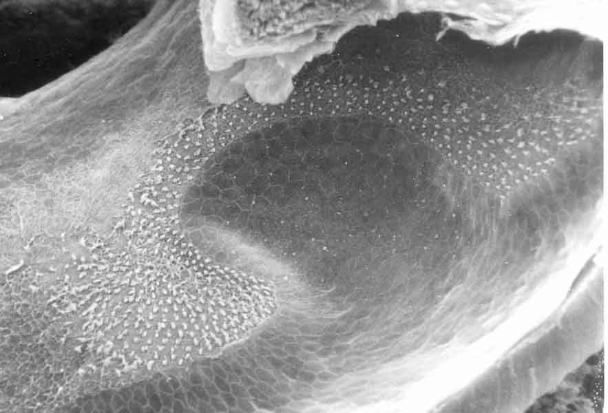
On the lower left is an AP from Sooglossus thomasetti , a species in the first branch (Sooglissidae) of the Hyloides limb.
The animal, caught on Mahe in the Seychelle Islands in 1985, was given to us by R.A. Nussbaum. On the lower right is an AP from
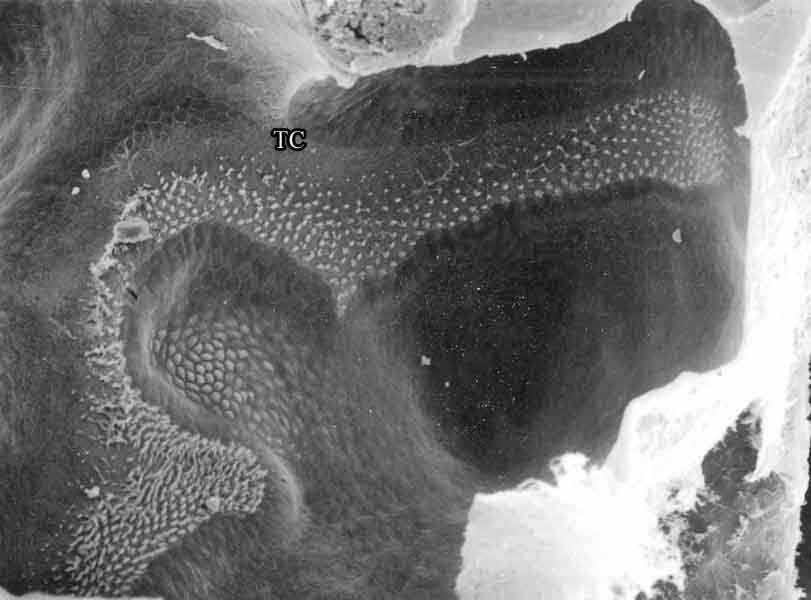
Gastrophryne olivacea, a species in the first branch (the Allodapanura) of the Ranoides limb.
The story that will emerge from these web pages is one of remarkable plasticity in the caudal patch of the Lalagobatrachia
(Figure),
with considerable evolutionary experimentation among pre-neobatrachian APs, and remarkable convergence among APs in the
Hyloides and Ranoides limbs.
| More APs from ancient anurans |
APs from mid-branch Hyloides |
APs from hylids | The puzzling bufonids part I |
The puzzling bufonids part II |
APs from Ranoides |
| APs on the Tree of Life |
APs on a Rana Tree |
APs on a Bufo Tree |
APs on an Eleutherodactylus Tree |
What does the caudal extension do? |
Discussion |
Calibrated Images
The following index provides links to micrographs from a 1984 paper (Lewis ER, On the frog amphibian papilla, Scanning Electron
Microscopy/1984/IV, pp. 1899-1913). They are shown with 600-pixel vertical resolution and with scale bars to indicate
the magnification. Each AP either was taken from the left ear, or its image was mirrored to make its orientation
consistent with the rest. The numerical order of the presentation reflects my thinking, in 1984, of the general pattern of
evolution of AP shape. In light of the new tree of life, that thinking has changed.
| 14. AP from the Texas toad |
15. AP from the Plains narrow-mouth toad |
16. AP from Strecker's chorus frog |
17. AP from the green treefrog |
18. AP from the wood frog |
19. AP from the Cascades frog |
Last updated 03/02/09