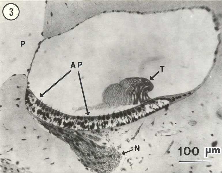
Light micrograph of a section through the amphibian papillar chamber of the American bullfrog, Rana catesbeiana. The section
is upside down; the amphibian papilla (the sensory surface), denoted AP, is on the ceiling of the chamber. The chamber itself is surrounded
largely by dense periotic connective tissue, and is innervated from above by the AP branch (N) of the VIIIth nerve. T denotes the tectorial
membrane (or "tectorium"), which has been distorted by the tissue preparation for light microscopy. The vertical spaces in the tectorium
are tubular, with one water-filled tube overlying each hair bundle. The contact membrane appears to be intact, lying between the amphibian
periotic canal (P) and the papillar chamber.
Last updated 01/22/09
|