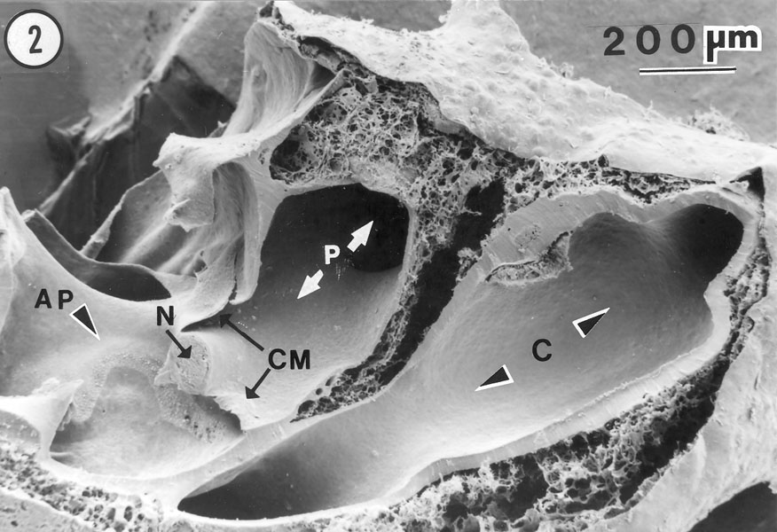
Scanning electron micrograph of the amphibian papilla (AP) and its surroundings from North America's largest toad, Bufo alvarius, the Colorado River toad.
Our view of the transected inner ear is from below (the ventral side). N denotes the AP branch of the VIIIth nerve. C denotes the confluence of the posterior-vertical
semicircular canal and the horizontal
semicircular canal. CM denotes the contact membranes, across which acoustic stimuli are presumed to leave the AP chamber and the neighboring region of the
saccular chamber. They pass out of the inner ear through the amphibian periotic canal (P). The sacculus has been removed, leaving just a small
part of its wall, in the vicinity of the AP. The letters N and AP are written on the ceiling of the opening between the saccular chamber
and the AP chamber. The tissue forming that ceiling also forms the floor of the utriculo-saccular foramen, which is clearly visible
here, just above the letters N and AP. Acoustic stimuli are presumed to enter the AP chamber from the saccular chamber, approximately in the
direction of the arrow pointing to the AP.
This piece of the toad's inner ear was mounted on a metal stub
for viewing in the electron microscope. Beneath the scale bar and figure number we are viewing the surface of the stub rather than tissue. The
thick walls around each chamber and canal are composed of dense periotic connective tissue. These are surrounded by spaces filled largely with water;
and the entire structure is enclosed in a thin layer of epithelium, part of which can be seen in the upper-right and in the lower-right corner.
Last updated 02/12/09
|