Chunlei Liu @ UC Berkeley
Medical Imaging, Brain Imaging and Cell Modulation
News
Victor Han: ISMRM 2020 I.I. Rabi Young Investigator Award Finalist
Congratulations!

Congratulations to Victor Han, Finalist for ISMRM I.I. Rabi Award
Victor Han (advisor: Prof. Chunlei Liu) was selected as a finalist for the International Society of Magnetic Resonance in Medicine (ISMRM) I.I. Rabi Young Investigator Award for original basic research. He was chosen for his paper entitled “Multiphoton Magnetic Resonance Imaging,” in which he developed a novel technique that excites multiphoton resonances to generate signal for MRI by using multiple magnetic field frequencies, none of which is near the Larmor frequency. Only the total energy absorbed by a spin must correspond to the Larmor frequency. In contrast, today’s MRI exclusively relies on single-photon excitation. He was named runner-up at the ISMRM annual conference in early August. Han will continue to develop his multiphoton technique and is exploring its applications in medicine and neuroscience as a part of his PhD dissertation research. The ISMRM is a multi-disciplinary nonprofit professional association that promotes innovation, development, and application of magnetic resonance techniques in medicine and biology throughout the world.
Trisha Shang: Paola S. Timiras Memorial Prize
Congratulations!

Congratulations to Trisha!
Paola S. Timiras Memorial Prize
Awarded to the top graduating senior in the Cell & Developmental Biology emphasis for high academic achievement and excellence of research in an honors project. Congratulations!
Lipid Oxidation Induced by RF Waves and Mediated by Ferritin Iron Causes Activation of Ferritin-Tagged Ion Channels
Miriam Hernández-Morales et al. Cell Reports 2020
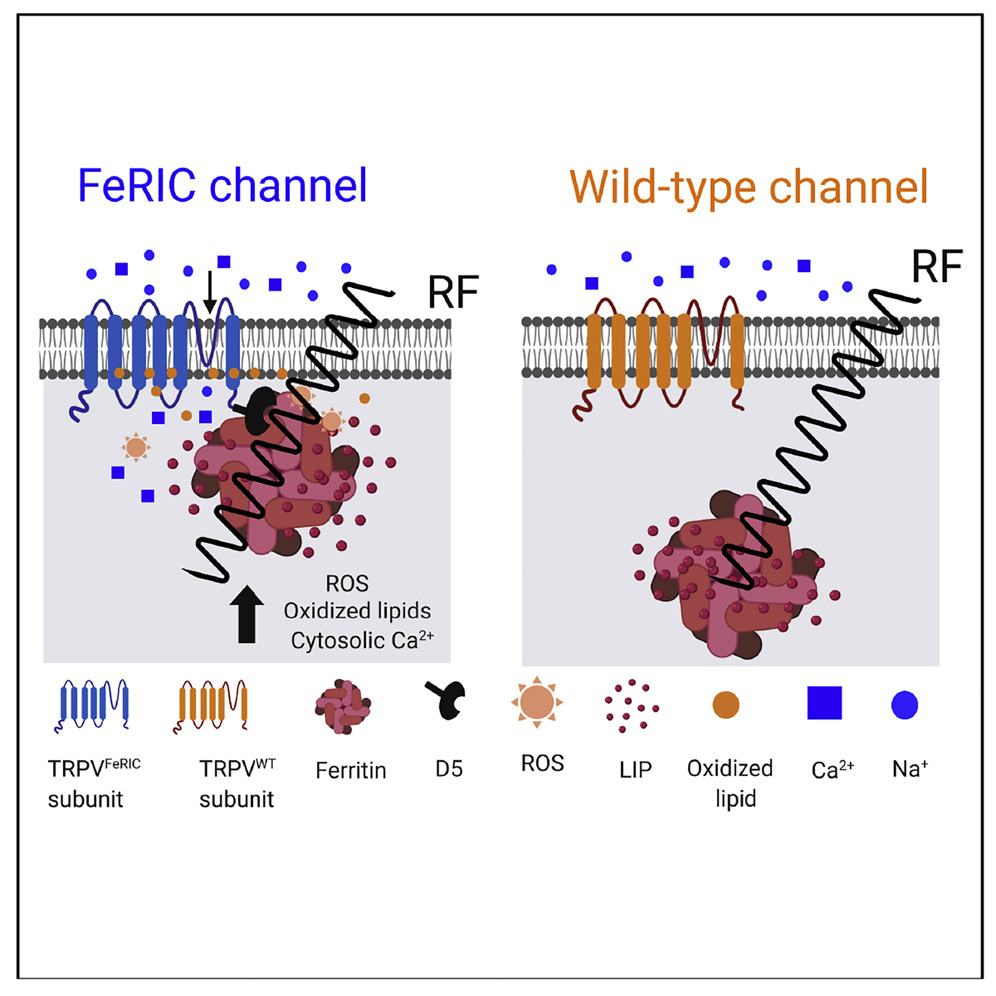
Lipid Oxidation Induced by RF Waves and Mediated by Ferritin Iron Causes Activation of Ferritin-Tagged Ion Channels
One approach to magnetogenetics uses radiofrequency (RF) waves to activate transient receptor potential channels (TRPV1 and TRPV4) that are coupled to cellular ferritins. The mechanisms underlying this effect are unclear and controversial. Theoretical calculations suggest that the heat produced by RF fields is likely orders of magnitude weaker than needed for channel activation. Using the FeRIC (Ferritin iron Redistribution to Ion Channels) system, we have uncovered a mechanism of activation of ferritin-tagged channels via a biochemical pathway initiated by RF disturbance of ferritin and mediated by ferritin-associated iron. We show that, in cells expressing TRPVFeRIC channels, RF increases the levels of the labile iron pool in a ferritin-dependent manner. Free iron participates in chemical reactions, producing reactive oxygen species and oxidized lipids that ultimately activate the TRPVFeRIC channels. This biochemical pathway predicts a similar RF-induced activation of other lipid-sensitive TRP channels and may guide future magnetogenetic designs.
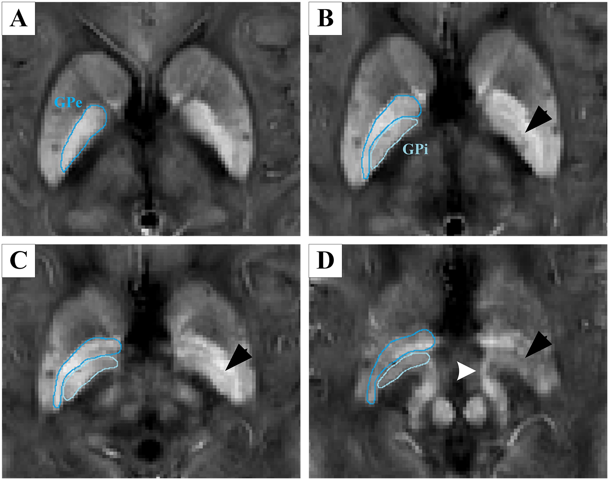
Precise targeting of the globus pallidus internus with quantitative susceptibility mapping for deep brain stimulation surgery
Hongjiang Wei et al. Journal of Neurosurgery, 2019
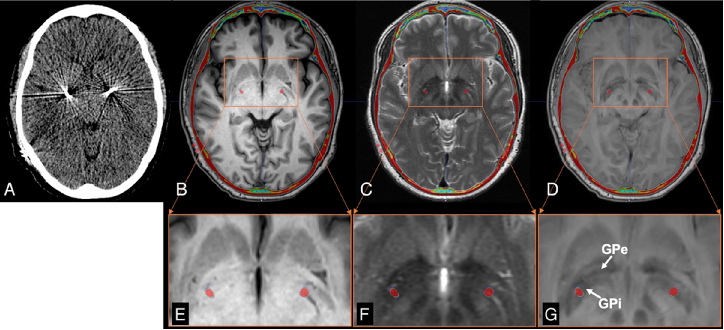
Precise targeting of the globus pallidus internus with quantitative susceptibility mapping for deep brain stimulation surgery
OBJECTIVE The goal of this study was to demonstrate the use of quantitative susceptibility mapping (QSM)–based images to precisely localize the globus pallidus internus (GPi) for deep brain stimulation (DBS) planning and to enhance postsurgical visualization of the DBS lead positions. METHODS Presurgical T1-weighted (T1w), T2-weighted (T2w), and QSM images as well as postsurgical CT images were obtained in 29 patients with Parkinson’s disease. To enhance the contrast within the GP, a hybrid contrast was created by linearly combining T1w and QSM images. Contrast-to-noise ratios (CNRs) of the GPi on T1w, T2w, QSM, and hybrid images were compared. The CNR differences were tested using the 1-way ANOVA method. The visualization of the DBS lead position was demonstrated by merging the postsurgical CT with presurgical MR images. RESULTS The hybrid images yield the best CNRs for GPi depiction and the visualization of the postsurgical DBS lead position was significantly improved. CONCLUSIONS QSM-based images allow for confident localization of borders of the GPi that is superior to T1w and T2w images. High-contrast hybrid images can be used for precisely directed DBS targeting, e.g., GPi DBS for the treatment of advanced Parkinson’s disease.
Temperature-activated ion channels in neural crest cells confer maternal fever–associated birth defects
Mary R. Hutson et al. Science Signaling 2017

Temperature-activated ion channels in neural crest cells confer maternal fever–associated birth defects
Birth defects of the heart and face are common, and most have no known genetic cause, suggesting a role for environmental factors. Maternal fever during the first trimester is an environmental risk factor linked to these defects. Neural crest cells are precursor populations essential to the development of both at-risk tissues. We report that two heat-activated transient receptor potential (TRP) ion channels, TRPV1 and TRPV4, were present in neural crest cells during critical windows of heart and face development. TRPV1 antagonists protected against the development of hyperthermia-induced defects in chick embryos. Treatment with chemical agonists of TRPV1 or TRPV4 replicated hyperthermia-induced birth defects in chick and zebrafish embryos. To test whether transient TRPV channel permeability in neural crest cells was sufficient to induce these defects, we engineered iron-binding modifications to TRPV1 and TRPV4 that enabled remote and noninvasive activation of these channels in specific cellular locations and at specific developmental times in chick embryos with radio-frequency electromagnetic fields. Transient stimulation of radio frequency–controlled TRP channels in neural crest cells replicated fever-associated defects in developing chick embryos. Our data provide a previously undescribed mechanism for congenital defects, whereby hyperthermia activates ion channels that negatively affect fetal development.
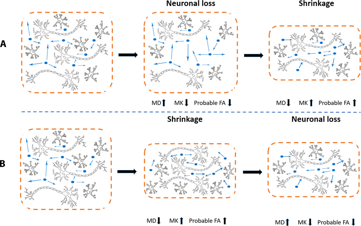
Microstructural alterations of cortical and deep gray matter over a season of high school football revealed by diffusion kurtosis imaging
Nan-Jie Gong et al, Neurobiology of Disease, 2018

Microstructural alterations of cortical and deep gray matter over a season of high school football revealed by diffusion kurtosis imaging
Objectives
To probe microstructural changes that are associated
with subconcussive head impact exposure in deep and
cortical gray matter of high school football players
over a single season.
Methods
Players underwent diffusion kurtosis imaging (DKI) and
quantitative susceptibility mapping (QSM) scans. Head
impact data was recorded. Association between parametric
changes and frequency of frontal head impact was
assessed.
Results
In deep gray matter, significant decreases in mean
kurtosis (MK) and increases in mean diffusivity (MD)
over the season were observed in the thalamus and
putamen. Correlations between changes in DKI metrics and
frequency of frontal impacts were observed in the
putamen and caudate. In cortical gray matter, decreases
in MK were observed in regions including the pars
triangularis and inferior parietal. In addition,
increases in MD were observed in the rostral middle
frontal cortices. Negative correlations between MK and
frequency of frontal impacts were observed in the
posterior part of the brain including the pericalcarine,
lingual and middle temporal cortices. Magnetic
susceptibility values exhibited no significant
difference or correlation, suggesting these diffusion
changes common within the group may not be associated
with iron-related mechanisms.
Implications for patient care
DKI may yield valuable biomarkers for evaluating the
severity of brain injuries associated with subconcussive
head impacts in contact sport athletes.
NMR in Biomedicine: 30 Year Celebration
Congratulations! Two papers from our lab were honored,
2018

In 2018, NMR in Biomedicine celebrates its 30th Birthday.
The articles included in this celebration issue have
been selected by the editors of NMR in Biomedicine. One
outstanding article from each of the thirty years that
the journal has published has been made freely
available.
Li, Wei, Alexandru V. Avram, Bing Wu, Xue Xiao, and
Chunlei Liu. "Integrated
Laplacian‐based phase unwrapping and background phase
removal for quantitative susceptibility mapping."
NMR in Biomedicine 27, no. 2 (2014): 219-227.
Guan, Xiaojun, Min Xuan, Quanquan Gu, Peiyu Huang,
Chunlei Liu, Nian Wang, Xiaojun Xu, Wei Luo, and Minming
Zhang. "Regionally
progressive accumulation of iron in Parkinson's
disease as measured by quantitative susceptibility
mapping." NMR in Biomedicine 30, no. 4 (2017):
e3489.
Chelsey Rodriguez: SURF Fellow 2017
Congratulations!
UC Berkeley Summer Undergraduate Research Fellowship
(SURF)
SURF L&S supports undergraduates in the College of Letters and Science to spend the summer doing concentrated research in preparation for a senior thesis or other major capstone research project. Fellows receive a summer stipend of approximately $4000, which is intended to cover basic living expenses for two months.
Hongjiang Wei: ISMRM Research Exchange Program 2017
Congratulations!

ISMRM Research Exchange Program 2017 Recipient Funded
by the ISMRM Research & Education Fund
Project
Title: Clinical Applications of Quantitative
Susceptibility Mapping in Human Knee Joints
Home Mentor – Chunlei Liu at UC Berkeley, CA, USA
Host Mentor – Fuhua Yan at Shanghai Jiao Tong
University, Shanghai, China .
Susceptibility tensor imaging and tractography of cartilage
Hongjiang Wei et al. Magnetic Resonance Medicine 2017
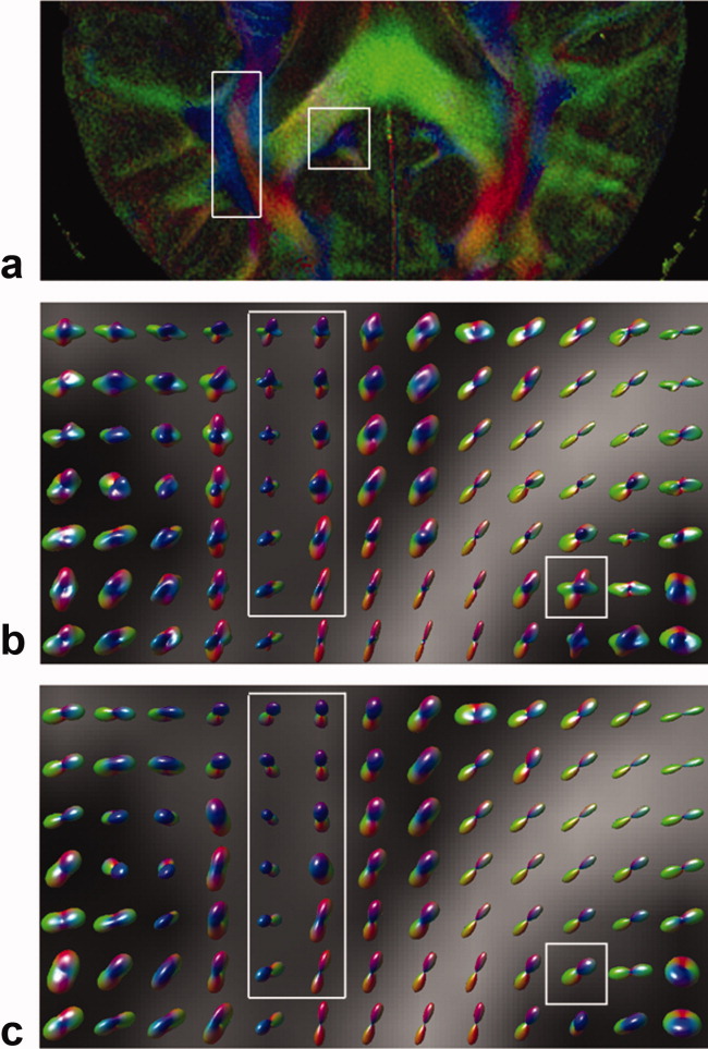
Susceptibility tensor imaging and tractography of collagen fibrils in the articular cartilage
Purpose
To investigate the B0 orientation-dependent magnetic
susceptibility of collagen fibrils within the articular
cartilage and to determine whether susceptibility tensor
imaging (STI) can detect the 3D collagen network within
cartilage.
Methods
Multiecho gradient echo datasets (100-μm isotropic
resolution) were acquired from fixed porcine articular
cartilage specimens at 9.4 T. The susceptibility tensor
was calculated using phase images acquired at 12 or 15
different orientations relative to B0. The
susceptibility anisotropy of the collagen fibril was
quantified and diffusion tensor imaging (DTI) was
compared against STI. 3D tractography was performed to
visualize and track the collagen fibrils with DTI and
STI.
Results
STI experiments showed the distinct and significant
anisotropic magnetic susceptibility of collagen fibrils
within the articular cartilage. STI can be used to
measure and quantify susceptibility anisotropy maps.
Furthermore, STI provides orientation information of the
underlying collagen network via 3D tractography.
Conclusion
The findings of this study demonstrate that STI can
characterize the orientation variation of collagen
fibrils where diffusion anisotropy fails. We believe
that STI could serve as a sensitive and noninvasive
marker to study the collagen fibrils microstructure.
Regionally progressive accumulation of iron in Parkinson's disease as measured by quantitative susceptibility mapping
Xiaojun Guan et al. NMR in Biomedicine 2016
Regionally progressive accumulation of iron in Parkinson's disease as measured by quantitative susceptibility mapping
The progression of Parkinson's disease (PD) seems to vary according to the disease stage, which greatly influences the management of PD patients. However, the underlying mechanism of progression in PD remains unclear. This study was designed to explore the progressive pattern of iron accumulation at different stages in PD patients. Sixty right‐handed PD patients and 40 normal controls were recruited. According to the disease stage, 45 patients with Hoehn–Yahr stage ≤ 2.5 and 15 patients with Hoehn–Yahr stage ≥ 3 were grouped into early‐stage PD (EPD) and late‐stage PD (LPD) groups, respectively. The iron content in the cardinal subcortical nuclei covering the cerebrum, cerebellum and midbrain was measured using quantitative susceptibility mapping (QSM). The substantia nigra pars compacta (SNc) showed significantly increased QSM values in the EPD patients compared with the controls. In the LPD patients, while the SNc continued to show increased QSM values compared with the controls and EPD patients, the regions showing increased QSM values spread to include the substantia nigra pars reticulata (SNr), red nucleus (RN) and globus pallidus (GP). Our data also indicated that iron deposition was more significant in the GP internal segment (GPi) than in the GP external segment. No other regions showed significant changes in QSM values among the groups. Therefore, we were able to confirm a regionally progressive pattern of iron accumulation in the different stages of PD, indicating that iron deposition in the SNc is affected exclusively in the early stages of the disease, while the SNr, RN and GP, and particularly the GPi segment, become involved in advanced stages of the disease. This is a preliminary study providing objective evidence of the iron‐related progression in PD.
Alzheimer's Disease: Microstructural Changes Precede Morphological Changes?
Nan-Jie Gong et al. Human Brain Mapping 2017
Differential microstructural and morphological abnormalities in mild cognitive impairment and Alzheimer's disease: Evidence from cortical and deep gray matter
A cohort of 18 patients with AD, 18 patients with amnestic mild cognitive impairment (MCI), and 18 normal controls underwent morphological and DKI MR imaging. Images were investigated using regions-of-interest-based analyses for deep gray matter and vertex-wise analyses for cortical gray matter. In deep gray matter, more regions showed DKI parametric abnormalities than atrophies at the early MCI stage. Mean kurtosis (MK) exhibited the largest number of significant abnormalities among all DKI metrics. At the later AD stage, diffusional abnormalities were observed in fewer regions than atrophies. In cortical gray matter, abnormalities in thickness were mainly in the medial and lateral temporal lobes, which fit the locations of known early pathological changes. Microstructural abnormalities were predominantly in the parietal and even frontal lobes, which fit the locations of known late pathological changes. In conclusion, MK can complement conventional diffusion metrics for detecting microstructural changes, especially in deep gray matter. This study also provides evidence supporting the notion that microstructural changes predate morphological changes.
Imaging Mouse Brain at 10-Micron Resolution
Hongjiang Wei, et al. NeuroImage 2016
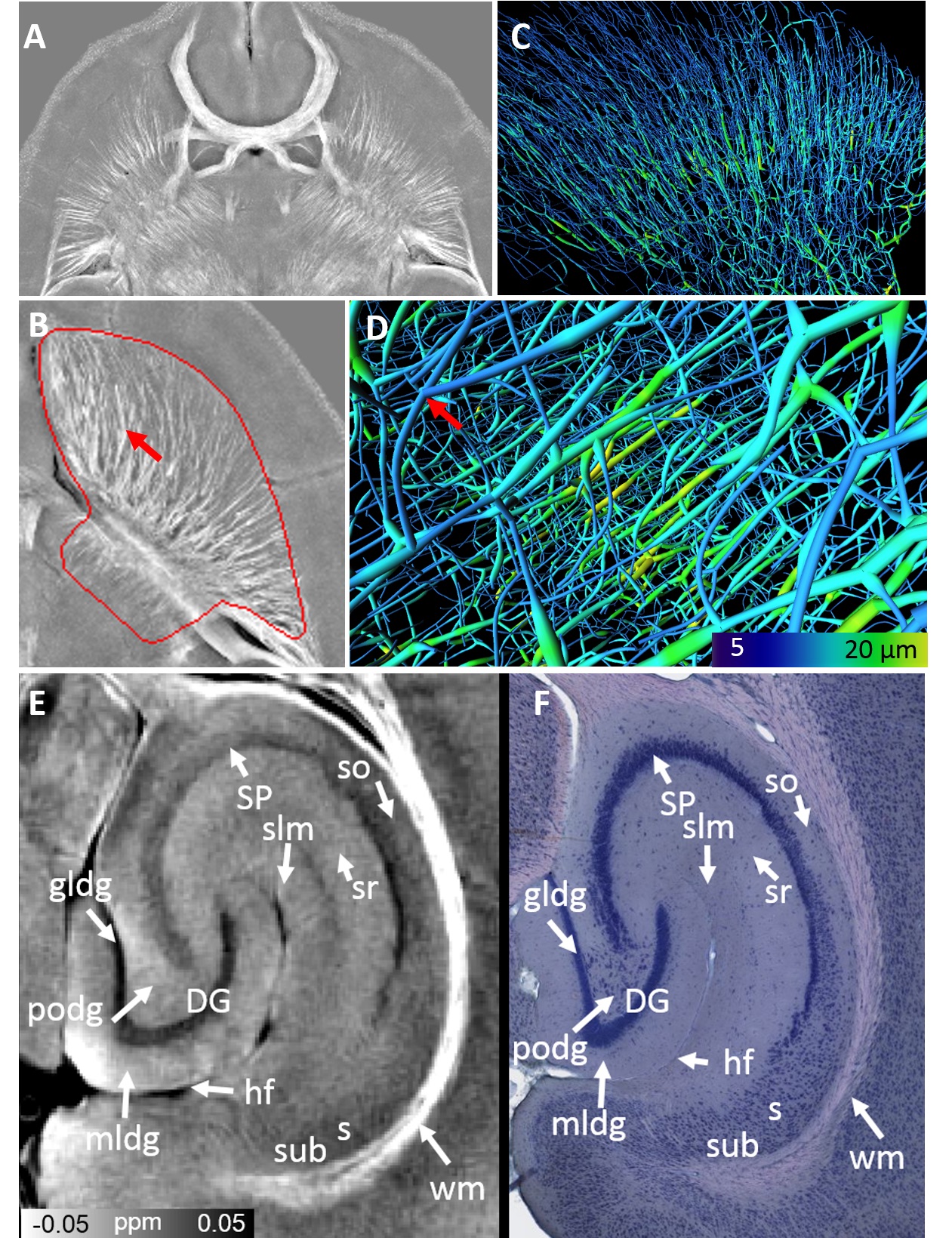
Imaging whole-brain cytoarchitecture of mouse with MRI-based quantitative susceptibility mapping
We present a new MRI method for imaging whole brain cytoarchitecture at 10-μm isotropic resolution non-destructively in 3D.
Cell layers are visualized in the olfactory bulb, eye ball, barrel cortex, hippocampus and cerebellum.
Axonal fiber trajectories are identified in both white matter and gray matter.
A network of medium spiny neurons are demonstrated in striatum.
STI of Brain, Kidney and Heart
Russell Dibb and Chunlei Liu, Magn Reson Med 2017
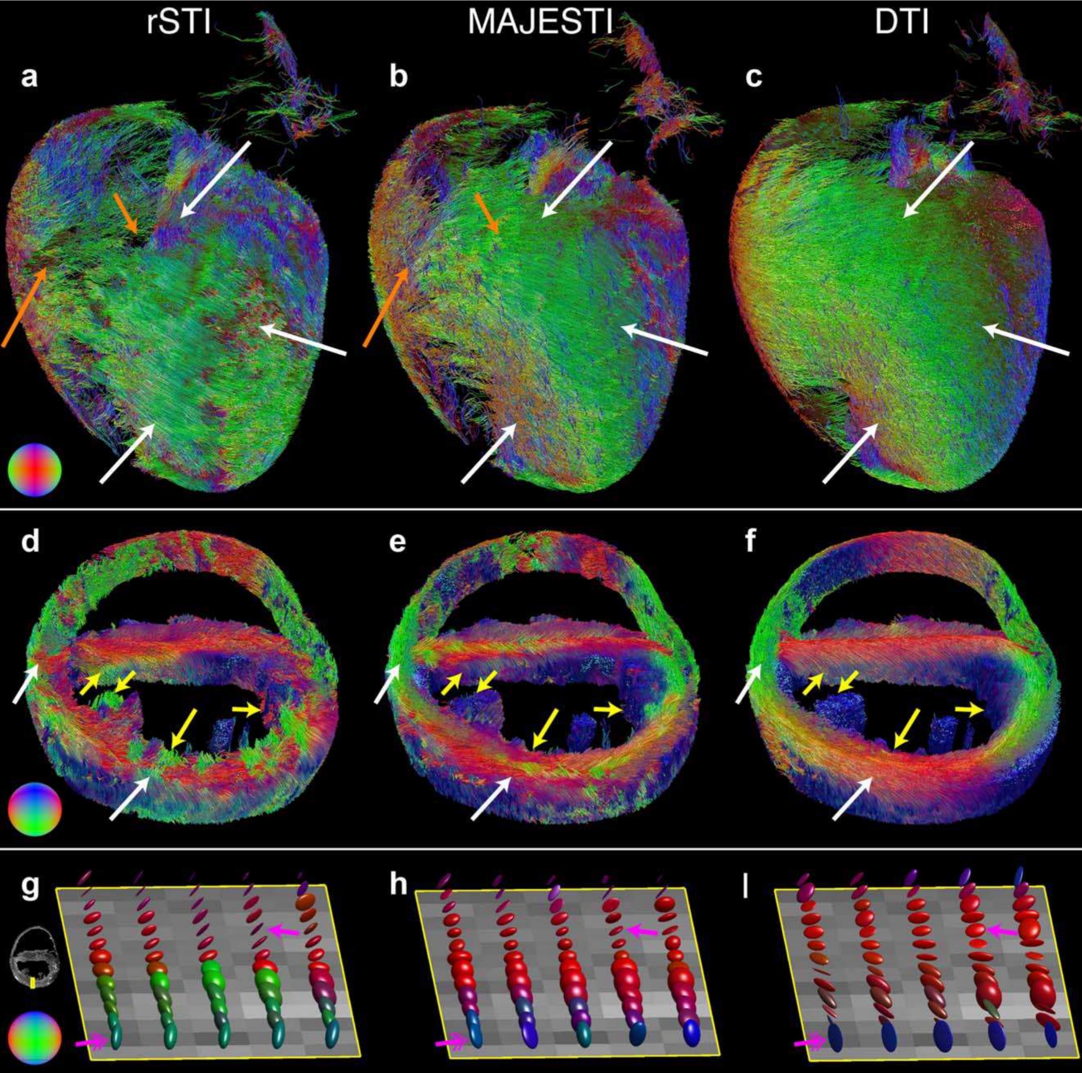
Joint eigenvector estimation from mutually anisotropic
tensors improves susceptibility tensor imaging of the
brain, kidney and heart
STI can probe tissue microstructure, but is limited by reconstruction artifacts because of absent phase information outside the tissue and noise. STI accuracy may be improved by estimating a joint eigenvector from mutually anisotropic susceptibility and relaxation tensors. Gradient-recalled echo image data were simulated using a numerical phantom and acquired from the ex vivo mouse brain, kidney, and heart. Susceptibility tensor data were reconstructed using STI, regularized STI, and the proposed algorithm of mutually anisotropic and joint eigenvector STI (MAJESTI). Fiber map and tractography results from each technique were compared with diffusion tensor data. MAJESTI estimation of the susceptibility tensors yields lower orientation errors for susceptibility-based fiber mapping and tractography in the intact brain, kidney, and heart.
Subtype of EAE with Neuronal Damage
Makoto Inoue, et al. Nature Neuroscience 2016
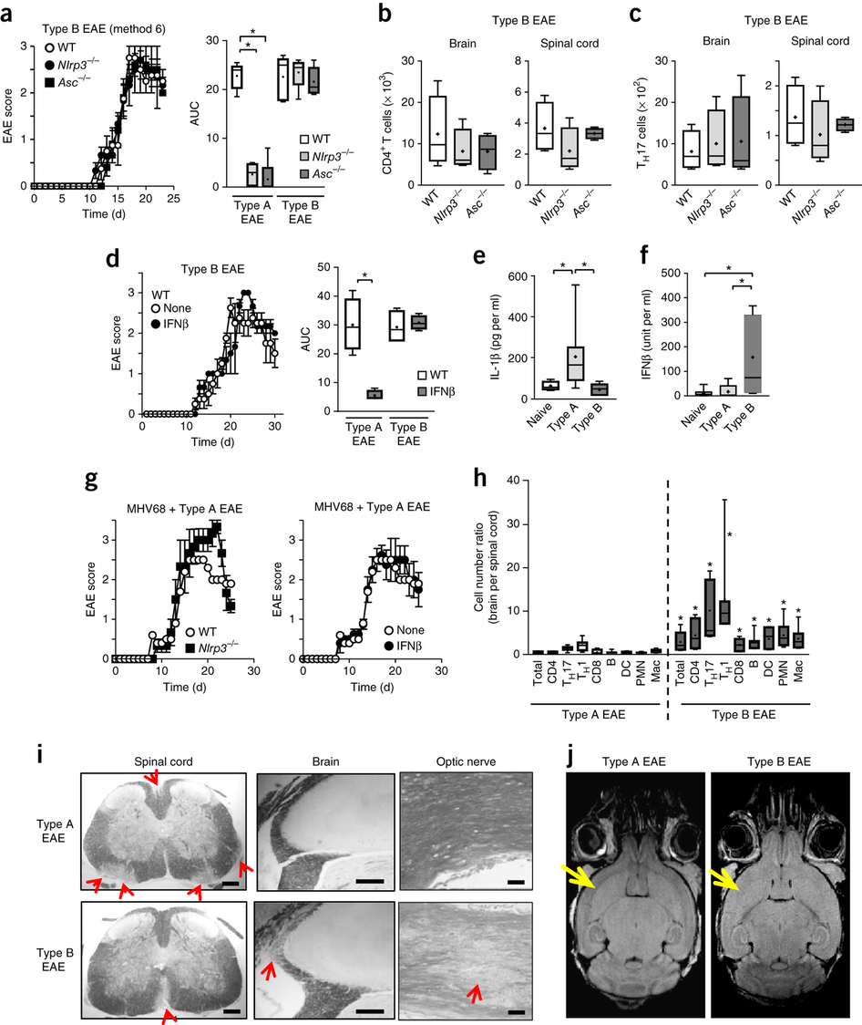
An interferon-β-resistant and NLRP3 inflammasome–independent subtype of EAE with neuronal damage
Inflammation induced by innate immunity influences the development of T cell–mediated autoimmunity in multiple sclerosis and its animal model, experimental autoimmune encephalomyelitis (EAE). We found that strong activation of innate immunity induced Nod-like receptor protein 3 (NLRP3) inflammasome–independent and interferon-β (IFNβ)-resistant EAE (termed type B EAE), whereas EAE induced by weak activation of innate immunity requires the NLRP3 inflammasome and is sensitive to IFNβ treatment. Instead, an alternative inflammatory mechanism, including membrane-bound lymphotoxin-β receptor (LTβR) and CXC chemokine receptor 2 (CXCR2), is involved in type B EAE development, and type B EAE is ameliorated by antagonizing these receptors. Relative expression of Ltbr and Cxcr2 genes was indeed enhanced in patients with IFNβ-resistant multiple sclerosis. Remission was minimal in type B EAE due to neuronal damages induced by semaphorin 6B upregulation on CD4+ T cells. Our data reveal a new inflammatory mechanism by which an IFNβ-resistant EAE subtype develops.
GDTI of Non-Gaussian Diffusion
Chunlei Liu, et al. Magn Reson Med 2004
Characterizing non-gaussian diffusion by using generalized diffusion tensors
Diffusion tensor imaging (DTI) is known to have a limited capability of resolving multiple fiber orientations within one voxel. This is mainly because the probability density function (PDF) for random spin displacement is non-Gaussian in the confining environment of biological tissues and, thus, the modeling of self-diffusion by a second-order tensor breaks down. The statistical property of a non-Gaussian diffusion process is characterized via the higher-order tensor (HOT) coefficients (variance, skewness, kurtosis etc.) by reconstructing the PDF of the random spin displacement. Those HOT coefficients can be determined by combining a series of complex diffusion-weighted measurements. The signal equation for an MR diffusion experiment was investigated theoretically by generalizing Fick's law to a higher-order partial differential equation (PDE) obtained via Kramers-Moyal expansion. A relationship has been derived between the HOT coefficients of the PDE and the higher-order cumulants of the random spin displacement. Monte-Carlo simulations of diffusion in a restricted environment with different geometrical shapes were performed, and the strengths and weaknesses of both HOT and established diffusion analysis techniques were investigated. The generalized diffusion tensor formalism is capable of accurately resolving the underlying spin displacement for complex geometrical structures, of which neither conventional DTI nor diffusion-weighted imaging at high angular resolution (HARD) is capable. The HOT method helps illuminate some of the restrictions that are characteristic of these other methods. Furthermore, a direct relationship between HOT and q-space is also established.
Magnetic Susceptibility of Knee Joint
Hongjiang Wei, et al. Magn Reson Med 2017
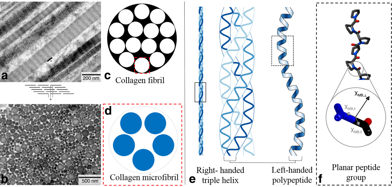
Investigating magnetic susceptibility of human knee joint at 7 tesla
A collagen tissue model was simulated and ex vivo animal cartilage experiments were conducted at 9.4 Tesla (T) to evaluate the B0 orientation-dependent magnetic susceptibility contrast observed in cartilage. Furthermore, nine volunteers (six healthy subjects without knee pain history and three patients with known knee injury, between 29 and 58 years old) were scanned using gradient-echo acquisitions on a high-field 7T MR scanner. Susceptibility values of different tissues were quantified and diseased cartilage and meniscus were compared against that of healthy volunteers. The arrangement of the collagen fibrils is significant, and likely the most dominant source of magnetic susceptibility anisotropy. Quantitative susceptibility mapping offers a means to characterize magnetic susceptibility properties of tissues in the knee joint. It is sensitive to collagen damage or degeneration and may be useful for evaluating the status of knee diseases, such as meniscal tears and cartilage disease.
Chunlei Liu, Ph.D.
Associate Professor
Department of Electrical Engineering and Computer Sciences,
and
Helen Wills Neuroscience Institute
University of California, Berkeley
Education
B.S. Physics, Peking University
M.S. Physics, University of North Carolina, Chapel Hill
M.S. Management Science and Engineering, Stanford University
Ph.D. Electrical Engineering, Stanford University
- Location: 505 Cory Hall, MC# 1770,
Berkeley, CA
- Tel: (510)664 7596